

Gross and Histopathological Lesions Associated with Platyhelminth Infections in Camels (Camelus dromedarius) at a Somali Meat Processing Facility
by
Abdirahman Barre 1,2,*,
Faez F. J. Abdullah 2,
Shafii A. Mohamed 3,
Hamza A. Hashi 1,
Krishnan N. Balakrishnan 4 and
Hodan I. Nageye 3
1
Faculty of Agriculture and Veterinary Medicine, Salaam University Mogadishu, Somalia
2
Department of Veterinary Clinical Studies, Faculty of Veterinary Medicine, University Putra Malaysia, 43400 Serdang, Selangor, Malaysia
3
Department of Infectious Disease, Faculty of Veterinary Medicine, Somali National University, Mogadishu, Somalia
4
Biotechnology Research Institute, University Malaysia Sabah, Kota Kinabalu, Sabah, Malaysia
*
Correspondence: idaajaaa007@gmail.com
Insights Anim. Sci. 2025, 2(1), 52–58.
https://doi.org/10.69917/ias.02.01-05
Received: April 18, 2025 /
Accepted: June 18, 2025 /
Published online: June 19, 2025
Abstract
This study aimed to investigate gross and histopathological liver lesions caused by Platyhelminthes in camels slaughtered at a meat processing facility in the Dynile District, Somalia. The investigations were conducted between April and October 2024. A total of 340 slaughtered camels were examined during the study. Among these, 200 camels (58.8%) were found to have liver lesions associated with Platyhelminthes based on both gross post-mortem examination and microscopic histopathological analysis. Gross pathological changes included hepatic swelling (34.5%), yellowish discoloration (27.5%), hemorrhagic lesions (11.5%), bile duct dilation (9.5%), necrosis (7.0%), cirrhosis (5.5%), and exudative lesions (4.5%). Histopathological analysis revealed frequent features such as tissue infiltration (25.5%), lymphocytic infiltration (18.1%), granuloma formation (13.5%), eosinophilic infiltrates (13.0%), necrosis (11.2%), abscess formation (8.3%), inflammation (7.1%), fibrosis (4.2%), and hepatocellular necrosis (2.4%). These findings highlight the pathological burden of platyhelminth infections in camels and underscore the potential zoonotic risks associated with their transmission.
Keywords:
Platyhelminthes; gross lesions; histopathology; slaughterhouse; camels
1. Introduction
Camels play a vital role in the livelihoods and culture of Somali communities, serving as a primary source of meat, milk, transportation, and income. Somalia possesses the largest camel population in Africa [1] and these animals are integral to both economic stability [2] and social traditions in the region. Despite their significance, camel health management remains a challenge due to limited veterinary services [3], inadequate disease monitoring systems, and the prolonged impact of civil conflict. These challenges contribute to underdiagnosis and poor control of infectious diseases affecting camel productivity and public health [4].
Among the various health issues affecting camels, parasitic infections caused by Platyhelminthes are of particular concern due to their prevalence and zoonotic potential [5]. These parasites can cause significant hepatic damage, leading to economic losses from condemned organs and decreased animal performance [6]. Infections such as Fasciola hepatica, Taenia saginata, and other trematodes are commonly found in livestock worldwide and pose public health risks [7], especially in areas with limited sanitation and veterinary oversight. Despite their importance, data on the pathological impacts of these parasites in camels, particularly in Somalia, remain scarce.
Therefore, this study was conducted to identify and characterize the gross and histopathological liver lesions caused by Platyhelminthes in camels slaughtered at the Deynile slaughterhouse in Mogadishu, Somalia. The findings are intended to contribute to the understanding of parasitic disease burden in camels and support future control and prevention strategies within a One Health framework.
2. Materials and Methods
2.1. Study Area
The study was conducted at a slaughterhouse located in Deynile District, Mogadishu, Somalia. Deynile is situated in the southeastern part of the Banadir region and is the smallest administrative district in Somalia, yet it has the largest population. The area is relatively stable, with favorable economic conditions and sufficient infrastructure to support the livestock trade and meat processing activities.
2.2. Study Design and Sampling
This cross-sectional study was conducted to assess the prevalence of gross and histopathological liver lesions associated with platyhelminth infections in camels slaughtered at a meat processing facility in the Deynile District of Somalia. A total of 360 camels were selected using a purposive sampling method. The sampled animals originated from various locations within Deynile District and included both sexes (80% male, 20% female). All studied camels were over seven years of age and exhibited good body condition, as determined through both ante-mortem and post-mortem evaluations.
2.3. Sample Collecion and Processing
Following ante-mortem inspection, post-mortem examination of slaughtered camels was conducted with a specific focus on the liver. Each liver underwent a thorough examination involving visual inspection, palpation, and incision to detect any gross pathological lesions. Observations were made regarding the size, appearance, and characteristics of the lesions, and the presence of Platyhelminth parasites during incision was also documented.
A total of 360 camels were examined, from which 200 liver tissue samples exhibiting gross lesions were selected for histopathological evaluation to assess pathological changes associated with parasitic infections.
For microscopic analysis, thin tissue sections were prepared from each liver sample. Due to the delicate nature of fresh tissue, chemical fixation was required to prevent distortion and facilitate the preparation of high-quality histological sections. Tissue fixation was performed using 10% neutral buffered formalin for 48 hours at room temperature. Following fixation, samples were processed using the paraffin embedding technique. This method involved dehydration through graded alcohols, clearing, and infiltration with molten paraffin wax, which solidified to support thin sectioning.
Paraffin-embedded tissue blocks were sectioned at a thickness of 5 µm using a rotary microtome. The sections were mounted on glass slides and stained with Hematoxylin and Eosin (H&E) to highlight normal and pathological structures. The stained slides were examined under a light microscope (Olympus CX23) using 10× and 40× objectives. Only qualitative histopathological assessment was performed.
All tissue samples were processed at the Department of Veterinary Pathology, Microbiology, and Parasitology, University of Nairobi. The sample collection and histological processing protocols were adapted from Yalew et al. [8], with appropriate quality control measures followed throughout the study.
2.4. Statistical Analysis
The collected data were organized and coded using Microsoft Excel and subsequently analyzed using SPSS software (version 20.0; IBM Corp., Armonk, NY, USA). Descriptive statistics were employed to determine the prevalence and distribution of gross and histopathological liver lesions associated with Platyhelminth infections.
3. Results and Discussion
A total of 340 slaughtered camels were examined during the study. Among these, 200 camels (58.8%) were found to have liver lesions associated with Platyhelminthes based on combined gross and microscopic examinations.
The gross pathological features observed during post-mortem inspection of camel livers are summarized in Table 1. The most commonly identified lesion was hepatic swelling, present in 34.5% of the cases, followed by yellowish discoloration in 27.5%, which may indicate hepatic dysfunction or bile pigment accumulation. These findings are consistent with previous studies by Yalew et al. [8] and Gebrehiwot [9], who also reported a high prevalence of liver swelling in camels and cattle slaughtered in various regions. The similarity in results suggests that liver swelling may be a common pathological outcome of Platyhelminthes infection across different livestock species and geographic areas.
The prevalence of lesions observed in the present study was notably higher than those reported by Yasine et al. [10] and Lopes et al [11], indicating a greater burden of Platyhelminthes infections in the study area. Notably, yellowish discoloration of the liver (27.5%) was one of the prominent gross lesions. This finding is relatively uncommon in Somalia but has been documented in similar studies conducted in East and South Arabian countries, including Iran [12] and Saudi Arabia [13], suggesting regional differences in parasite exposure and pathology.
Other notable gross lesions included hemorrhagic lesions (11.5%), bile duct dilation (9.5%), necrotic foci (7.0%), cirrhotic changes (5.5%), and exudative cirrhosis (4.5%). These alterations reflect the pathological impact of Platyhelminth infections on liver morphology and function.
Table 1. Gross pathological lesions observed during post-mortem examination (n = 200).
| Gross features | Frequencies | Percentage (%) |
|---|---|---|
| Liver Swollen | 69 | 34.5 |
| Yellowish discoloration | 55 | 27.5 |
| Haemorrhagic lesions | 23 | 11.5 |
| Bile duct dilation | 19 | 9.5 |
| Necrosis | 14 | 7.0 |
| Cirrhosis | 11 | 5.5 |
| Exudates | 9 | 4.5 |
Histopathological examination of camel liver samples revealed a variety of lesions associated with Platyhelminth infections Table 2. The most frequently observed feature was tissue infiltration, detected in 51 cases (25.5%). This was followed by lymphocytic infiltration in 37 cases (18.1%) and granuloma formation in 29 cases (13.5%), indicating a chronic inflammatory response to parasitic presence. Such observations support the hypothesis that Platyhelminthes infections elicit a significant immunopathological response, contributing to both hepatic damage and systemic health implications.
Table 2. Microscopic lesions observed in post-mortem histopathological examination (n = 200).
| Histopathological features | Frequencies | Percentage (%) |
|---|---|---|
| Tissue Infiltration | 51 | 25.5 |
| Lymphocytic infiltration | 37 | 18.1 |
| Granuloma formation | 29 | 13.5 |
| Necrosis | 21 | 11.2 |
| Abscess formation | 17 | 8.3 |
| Inflammation | 13 | 7.1 |
| Infiltration | 11 | 5.5 |
| Fibrosis | 9 | 4.2 |
| Granulomas | 7 | 3.2 |
| Hepatocellular necrosis | 5 | 2.4 |
Moreover, tissue and lymphocytic infiltration can lead to a variety of histopathological conditions, including autoimmune disorders, where the immune system attacks healthy cells. These infiltrations have been associated with vascular inflammation, malignant lymphomas, and dysregulated immune responses. In the context of hepatic pathology, such infiltrations suggest an active immunological role in liver damage, particularly through mechanisms such as necrosis. The presence of both tissue and lymphocyte infiltration in this study indicates ongoing inflammatory and immune-mediated processes contributing to hepatic injury.
Other notable lesions included necrosis in 21 samples (11.2%), abscess formation in 17 (8.3%), and general inflammatory changes in 13 samples (7.1%). Additional findings comprised non-specific infiltration (5.5%), fibrosis (4.2%), well-formed granulomas (3.2%), and hepatocellular necrosis in 5 cases (2.4%), which likely resulted from direct tissue damage due to parasite migration through hepatic parenchyma.
The prevalence of granulomas and necrosis observed in the present study exceeds the levels reported by Sulieman et al. [14] and Manga-González and Ferreras [15] yet remains lower than those documented by Islam [16], who investigated the epidemiology and hepatic pathology of Fascioliasis, a parasitic infection caused by Platyhelminthes.
The prevalence of inflammation and fibrosis in current study was higher than those reported by Ngetich [17] in Kenya. Although both inflammation and fibrosis are components of tissue repair and regeneration, prolonged or excessive inflammation can lead to pathological fibrosis. This process involves the accumulation of fibrous connective tissue in and around damaged areas, which may result in scarring, organ dysfunction, or even failure. Chronic inflammation, triggered by parasitic infections, autoimmune responses, toxins, or trauma, is a key driver of this process.
Additionally, three less common microscopic lesions were identified in this study. Hepatocellular necrosis, indicative of tissue destruction due to parasitic migration, was observed alongside eosinophilic infiltration and bile duct swelling or dilation. These findings are consistent with those reported by others [18, 19], who documented similar hepatic lesions in animals infected with Platyhelminthes. Such pathological changes underscore the complex impact of parasitic infections on hepatic architecture and highlight the urgent need for improved control and prevention strategies in endemic regions.
Representative gross lesions caused by Platyhelminth infections are illustrated in Figure 1. These include hemorrhagic lesions, characterized by tissue destruction and localized bleeding; bile duct dilatation and cyst formation associated with chronic infection; and fibrotic changes with granuloma formation surrounding parasitic structures or eggs. In advanced cases, chronic infections may progress to cirrhosis, resulting in a firm, nodular liver.
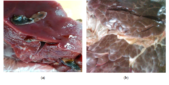
Figure 1. Gross pathological features of camel liver affected by Platyhelminth infection: (a) cyst and granuloma formation visible on the hepatic surface; (b) Firm, nodular liver indicative of cirrhosis associated with chronic parasitic infection.
A detailed histological section of the liver (Figure 2) shows the presence of granulomas surrounding parasitic structures, likely flatworms such as Fasciola hepatica or their eggs, embedded within the liver parenchyma. Prominent eosinophilic infiltrates and areas of necrosis are evident, indicating tissue damage associated with the host immune response. Periportal fibrosis and bile duct alterations are also observed around affected regions. Inflammatory cell infiltration is clearly visible, with numerous eosinophils, lymphocytes, and macrophages localized near the lesions.

Figure 2. Histopathological features of camel liver infected with Platyhelminthes, showing (A) bile duct hyperplasia, (B) lymphocytic infiltration, (C) hepatic necrosis, and (D) hemorrhagic areas adjacent to bile ducts.
5. Conclusions
The present study demonstrates a significant burden of hepatic pathology associated with Platyhelminth infections in slaughtered camels, as evidenced by a high prevalence of both gross and microscopic lesions. Gross pathological findings such as liver swelling, yellowish discoloration, and bile duct dilatation were commonly observed, while histopathological examination revealed frequent occurrences of tissue infiltration, lymphocytic infiltration, granuloma formation, necrosis, and fibrosis. Less common but notable lesions included hepatocellular necrosis, eosinophilic infiltration, and bile duct hyperplasia, reflecting chronic parasitic damage. These findings emphasize the substantial impact of Platyhelminth infections on camel health and liver function. The study underscores the need for routine parasitological surveillance, improved antemortem diagnosis, and the implementation of targeted control measures to reduce the prevalence and pathological consequences of these infections in endemic regions.
Author Contributions
Conceptualization, methodology, A.B., F.F.J.A. and S.A.M.; resources, H.A.H., and H.I.N.; data curation, A.B. and F.F.J.A.; writing—original draft preparation, A.B. F.F.J.A. and S.A.M.; writ-ing—review and editing, K.N.B. and H.I.N. All authors have read and agreed to the published version of the manuscript.
Institutional Review Board Statement
This study was approved by the Salaam University and Somali National University, Faculty of Veterinary Medicine Committee.
Acknowledgments
The author sincerely would like to appreciate Some-meat Slaughterhouse (their facility to conduct the study). Salaam University and University of Nairobi Kenya University College of Veterinary Medicine Department of Pathology, Microbiology and Parasitology for provision of the laboratory and other facilities.
Conflicts of Interest
The authors declare no conflict of interest.
References
- Barre, A. Prevalence of hemorrhagic septicemia in dromedary camel (Camelus dromedarius) of some selected farms at Benadir region, Somalia. J. Istanbul Vet. Sci. 2023, 7 (1), 8-14. [Google Scholar] [CrossRef]
- Abri, M. A. A.; Faye, B. Genetic improvement in dromedary camels: challenges and opportunities. Front. Genet. 2019, 10, 167. [Google Scholar] [CrossRef]
- Zhokhov, A. E. Metacercariae of trematodes (Plathelminthes: Trematoda) of Garra dembecha (Actinopterygii: Cyprinidae) from Lake Tana, Ethiopia. Zoosyst. Ross. 2012, 21 (2), 193-203. [Google Scholar] [CrossRef]
- Cornwell, M. S.; Dodd, C. C. The role of Veterinary Services in areas of conflict. Rev. Sci. Tech. 2020, 39 (2), 451-460. [Google Scholar] [CrossRef]
- Bedossa, P.; Poitou, C.; Veyrie, N.; Bouillot, J. L.; Basdevant, A.; Paradis, V.; Clément, K. Histopathological algorithm and scoring system for evaluation of liver lesions in morbidly obese patients. Hepatology 2012, 56 (5), 1751-1759. [Google Scholar] [CrossRef]
- Strydom, T.; Lavan, R. P.; Torres, S.; Heaney, K. The economic impact of parasitism from nematodes, trematodes and ticks on beef cattle production. Animals 2023, 13, 1599. [Google Scholar] [CrossRef]
- Kiani, B.; Budke, C. M.; Shams Abadi, E.; Hashtarkhani, S.; Raouf Rahmati, A.; AkbarPour, M.; Zarean, M.; Hosseini Farash, B. R.; Kiani, F.; Moghaddas, E. Evaluation of zoonotic platyhelminthe infections identified in slaughtered livestock in Iran, 2015-2019. BMC Vet. Res. 2021, 17 (1), 185. [Google Scholar] [CrossRef]
- Yalew, K. W.; Awol, N.; Tsegay, Y.; Abraha, H. A study on gross and histopathological pulmonary lesions of cattle slaughtered at Abergelle Abattoir, Mekelle, Tigray, Ethiopia. J. Vet. Med. Anim. Health 2018, 10 (6), 148-152. [Google Scholar] [CrossRef]
- Gebrehiwot, T.; Berihu, K.; Birhanu, H.; Verma, P. C. Study on gross pulmonary lesions in lungs of slaughtered animals and their economic importance in Tigray, Ethiopia. Momona Ethiop. J. Sci. 2015, 7 (1), 46-54. [Google Scholar] [CrossRef]
- Yasine, A.; Daba, M.; Ashenafi, H.; Geldhof, P.; Van Brantegem, L.; Vercauteren, G.; Govaere, J. Tissue (re)distribution of Trypanosoma equiperdum in venereal infected and blood transfused horses. Vet. Parasitol. 2019, 268, 87-97. [Google Scholar] [CrossRef]
- Lopes, C. E.; Xavier, F. G.; Nicolino, R. R.; Cordeiro, L. F.; Rezende, L. C.; Lopes, M. C.; Ecco, R. Pathological findings and differential diagnoses of lymph node diseases in slaughtered cattle in Brazil: a study of 2000 samples. Vet. Pathol. 2024, 61 (6), 952-964. [Google Scholar] [CrossRef]
- Sazmand, A.; Joachim, A. Parasitic diseases of camels in Iran (1931-2017) - a literature review. Parasite 2017, 24, 21. [Google Scholar] [CrossRef]
- Alanazi, A. D.; Nguyen, V. L.; Alyousif, M. S.; Manoj, R. R.; Alouffi, A. S.; Donato, R.; Otranto, D. Ticks and associated pathogens in camels (Camelus dromedarius) from Riyadh Province, Saudi Arabia. Parasites Vectors 2020, 13, 1-9. [Google Scholar] [CrossRef]
- Sulieman, Y.; Ibrahim, S. O.; Eltayeb, R. E.; Pengsakul, T.; Afifi, A.; Zakaria, M. A.; Khairala, M. A. Gastrointestinal helminth parasites of ruminants slaughtered in Shendi abattoir, River Nile State, Sudan. J. Coast. Life Med. 2017, 5 (6), 249-253. [Google Scholar] [CrossRef]
- Manga-González, M. Y.; Ferreras, M. C. Dicrocoeliidae family: major species causing veterinary diseases. In Digenetic Trematodes; Springer: 2019; pp 279-319. [Google Scholar] [CrossRef]
- Islam, K. M. Epidemiological and pathological studies of fascioliasis in goats in Sylhet region of Bangladesh and investigation on effects of different liver tonic on pathology of fascioliasis; Doctoral dissertation, University of Rajshahi: Bangladesh, 2015. [Google Scholar]
- Ngetich, E. C. Prevalence and the effect of parasites on haematological parameters of livestock in the upper Kerio Valley, Kenya; Doctoral dissertation, University of Eldoret: Kenya, 2019. [Google Scholar]
- Herrera-Torres, G.; Ruiz-Campillo, M. T.; Bautista, M. J.; Martínez-Moreno, F. J.; Zafra, R.; Buffoni, L.; Rufino-Moya, P. J.; Martínez-Moreno, Á.; Molina-Hernández, V.; Pérez, J. Liver histopathological and immunohistochemical evaluation from Fasciola hepatica experimentally infected and reinfected sheep. Animals 2024, 14, 1833. [Google Scholar] [CrossRef]
- Ali, H.; Alhamrashdi, A.; El-Neweshy, M.; Asi, M. N.; Johnson, E. H.; Elshafie, E. I. An abattoir based-survey of helminthic liver infections and associated pathological lesions in sheep in the sultanate of Oman. Pak. Vet. J. 2023, 43, 194-198. [Google Scholar]
Publisher’s Note: The views expressed in all publications, including statements, opinions, and data, are solely those of the individual author(s) and contributor(s) and do not necessarily reflect the views of Insights Academic Publishing (IAP) and/or its editor(s). IAP and/or its editor(s) are not responsible for any injury to persons or damage to property resulting from the ideas, methods, instructions, or products referenced in the content.
Copyright: © 2025 by the authors.
License: This article is published under the Creative Commons Attribution 4.0 International.CC BY 4.0
Publisher: Insights Academic Publishing (IAP), Lahore, Pakistan.