

Single-Cell Omics in Animal Reproductive Biology: A Cellular and Molecular Atlas Across Developmental Stages
byAbstract
The cutting-edge technologies of single-cell omics have revolutionized the study of animal reproduction, enabling the visualization of cellular and molecular domains in organisms during various growth stages. This review presents a conceptual framework grounded in the latest developments from single-cell RNA sequencing (scRNA-seq), ATAC sequencing (scATAC-seq), spatial transcriptomics, and multi-omics approaches applied to the reproductive organs of livestock and model species. Technologies survey molecular types of the cell, the course of transcription, chromatin accessibility, and regulatory networks for specific tissues in the development of gonads, gametogenesis, and embryo formation. In comparative analysis, both common and species-specific characteristics have been identified in models of cattle, pigs, goats, sheep, and avian species. This has made them a valuable resource for the development of trait-based breeding protocols. We discuss how single-cell data are being integrated with genomic selection frameworks to enhance fertility prediction and assisted reproduction strategies, leading to improvements in artificial insemination and reproductive disease modelling. The rapid evolution of protocols and tools has enabled the livestock science field to expand the use of these technologies, despite technical and computational challenges such as tissue dissociation bias, genome annotation gaps, and data complexity. The livestock science sector, therefore, is experiencing the broad adoption of single-cell differentials not just in the transformation of reproductive science but also in practical decision-making guidance for the new generation of animal breeding and fertility management.
Keywords: Single-cell RNA-seq; livestock reproduction; spatial transcriptomics; fertility biomarkers; reproductive genomics1. Introduction
Livestock productivity and evolutionary biology are highly dependent on reproductive success, which is why the latter is considered a cornerstone of both [1-3]. Traditional transcriptomics and bulk tissue analyses have significantly contributed to our understanding of reproductive organ function; however, they have often overlooked cellular heterogeneity, developmental trajectories, and lineage-specific regulatory mechanisms. The advent of single-cell omics technologies, particularly single-cell RNA sequencing (scRNA-seq), single-cell ATAC sequencing (scATAC-seq), and spatial transcriptomics, is fundamentally changing the field of reproductive biology by providing previously unattainable insights [4-6].
In mammals, reproduction is a multifaceted process involving various specialized cell types that undergo dynamic changes over the course of development, from primordial germ cells to mature gametes and embryonic formations. To enhance fertility techniques, identify biomarkers of reproductive disorders, and inform breeding strategies, understanding cellular and molecular identities, transitions, and regulatory programs is indeed essential [7-11]. Although early single-cell studies have demonstrated the mapping of testicular germ cell progression, the case of primary ovarian folliculogenesis and embryonic lineage commitment in humans and mice is similar [12-15]. Recent efforts are extending these insights to livestock species, including cattle, pigs, goats, and chickens [11, 16-20].
The use of single-cell omics technologies in livestock reproduction research holds not only immense biological relevance but also translational value [5,16,21-23]. For example, this enables the precise mapping of gene expression and chromatin landscapes. Pioneering single-cell initiatives have resolved cell types and their regulatory states across tissues, enabling integrated views of expression, chromatin accessibility, and regulatory networks. New strategies, such as improved semen selection, oocyte maturation protocols, and embryo transfer systems, will be employed [12,24-26]. Besides, single-cell profiling not only provides a means for identifying species- and breed-specific regulatory elements but also enables the possible integration of these regulatory elements into genomic selection programs, which ultimately leads to improvements in reproductive efficiency and resilience [9,27-29].
This review encompasses the current single-cell omics research in animal reproductive biology, with a primary focus on the technical applications in understanding gonadal development, gametogenesis, and embryo formation. We first compare the discoveries made in different species, then mention various new tools and methods, address problems that can only occur in livestock, and finally present a scenario of how these might influence the cropping and fertility management sector in the years to come.
2. Overview of Single-Cell Omics Technologies in Reproductive Biology
Over the last decade, single-cell technologies have experienced unprecedented growth, enabling the assessment of gene expression, chromatin accessibility, spatial localisation, and molecular interactions at the individual cell level [17,30-33]. The inventions of these tools have led to a paradigm shift in cellular diversity and dynamics research, particularly in complex and heterogeneous systems such as the reproductive organs. This section discusses the essential single-cell omics platforms currently used or anticipated to be beneficial for the future progress of reproductive biology in model organisms and livestock species [11,25,34-36].
2.1. Single-Cell RNA Sequencing (scRNA-seq)
One of the most widely used single-cell methods to date is single-cell RNA sequencing (scRNA-seq). It is the only method that simultaneously maps the transcriptome of thousands of cells and reveals the states of the cells, lineage trajectories, and transcriptional programs [21,24,37,38]. In the case of reproductive tissues, scRNA-seq was the technique used to reconstruct germ-cell progress [39], the development of follicles [12] that include Sertoli–germ-cell [37,40] interactions and cell fate of the embryos [41]. Platforms like 10x Genomics Chromium, Drop-seq, and Smart-seq2 vary in sensitivity, throughput, and cost, offering flexibility depending on the biological question and tissue type.
2.2. Single-Cell ATAC Sequencing (scATAC-seq)
While single-cell RNA sequencing profiles the expression of genes, single-cell ATAC sequencing provides a demonstration of chromatin alteration in each cell. This method examines chromatin openness, enhancers, and potential transcription-factor binding sites to identify the various gene-regulatory mechanisms at play in a particular cell [4,5,21,30]. In the context of reproduction, scATAC-seq has been utilised to elucidate the regulatory features during the processes of sperm-cell differentiation and egg-cell maturation, as well as chromatin-network restructuring in early embryos [1,6,42]. The most used instruments in the analysis and integration of these datasets are SnapATAC, ArchR, and Signac.
2.3. Spatial Transcriptomics
The primary disadvantage of dissociative single-cell methods is that they cannot retain spatial information. This is achieved through a spatial-transcriptomics approach, which enables the quantification of gene expression at specific coordinates without compromising the tissue architecture [17,18,32]. This method has been utilized in reproductive biology to track the distribution of spermatogonial stem cells in the seminiferous tubules [5,43,44] or to analyze the signaling of the oocyte to the granulosa cell in the follicles. Other recent devices, such as Visium (10x Genomics), MERFISH, and Slide-seq, have enabled the development of high-throughput, spatially resolved transcriptomic atlases.
2.4. Multi-Omics and Integrative Platforms
With the help of recent breakthroughs, it is now possible to provide multiple-omics profiles simultaneously from a single cell — such as gene expression (RNA), chromatin accessibility (ATAC), and protein abundance (CITE-seq, REAP-seq). The use of multi-omics strategies is classically necessary for the precise depiction of reproductive cell states and for recognising the regulatory events that are not captured in transcriptomics data [38,45,46]. Furthermore, computational platforms such as Seurat v4, MOFA+, and Harmony facilitate the integration of modal and time-point analysis.
Table 1. Summary of single-cell technologies used in reproductive biology.
| Technology | Resolution | Modality | Application in Reproduction | Key Advantages | Limitations |
|---|---|---|---|---|---|
| 10x Genomics scRNA-seq | 1 cell | mRNA transcripts | Gametogenesis, folliculogenesis, embryo profiling | High throughput, widely used | Dropout effect, ambient RNA |
| Smart-seq2 | 1 cell | Full-length mRNA | Low-input oocytes, rare cell types | High sensitivity, full-length coverage | Low throughput, costly |
| scATAC-seq | 1 cell | Chromatin accessibility | Germline enhancer discovery, TF binding dynamics | Identifies regulatory elements | Sparse data, high noise |
| 10x Multiome | 1 cell | RNA + ATAC | Integrated regulatory map in gametes and embryos | Multi-modal insight | High cost, complex analysis |
| CITE-seq | 1 cell | RNA + surface proteins | Immune profiling during implantation | Combines transcriptomics and proteomics | Antibody availability |
| Slide-seq | 10 µm | Spatial transcriptomics | Testis and ovary zonation | High spatial resolution | Limited transcriptome depth |
| Visium (10x) | 50 µm | Spatial transcriptomics | Uterine receptivity, follicular architecture | Whole-transcriptome, good tissue coverage | Moderate resolution |
The table presents several popular single-cell platforms, highlighting their key technical features, including resolution, captured modalities, applications in reproductive tissues, and notable advantages and limitations. These technologies constitute the core experimental toolkit for single-cell studies in reproductive biology across multiple species.
3. Single-Cell Transcriptomics in Reproductive Organs
Understanding the cellular composition and transcriptional dynamics of reproductive organs is crucial to deciphering the mechanisms of fertility, gametogenesis, and embryonic development. Single-cell RNA sequencing (scRNA-seq) is a transformative tool that has enabled researchers to clarify previously unidentified cell types, lineage trajectories, and stage-specific transcriptional programs in the testis, ovary, uterus, and placenta. This section integrates the relevant findings from scRNA-seq studies conducted on various reproductive tissues and animal species.
3.1. Testis: Dissecting Spermatogenesis at Single-Cell Resolution
The male mammalian reproductive system is highly intricate and comprises germ cells at its centre, supported by a somatic niche consisting of Sertoli, Leydig, and peritubular myoid cells. Single-cell RNA sequencing studies in mice [14,47-50], humans [35,41,51], and livestock species, such as cattle [16,17] and pigs [11,36], have substantially proved the existence of both conserved and species-specific patterns of spermatogenic development. UMAP and pseudotime analyses have revealed the discrete transitions of elongating spermatids from spermatogonial stem cells (SSCs), with the expression signatures of PLZF, KIT, SYCP1, and PRM1 that were all very apparent at each state.
Sertoli cells exhibit dynamic transcriptional alterations through interactions with germ cells, thereby activating genes related to phagocytosis, junctional remodelling, and hormonal regulation [24,40,52,53]. Within adult testis atlases (bovine and porcine), analyses revealed previously unrecognised heterogeneity within SSC populations, challenging the notion of linear differentiation.
3.2. Ovary: Folliculogenesis and Oocyte–Somatic Interactions
The intricate interplay of oocytes and granulosa cells governs
follicular development in the ovary. scRNA-seq profiling of fetal and
adult ovaries in mice [54,55], goats [18,52], and pigs [11,36] has been instrumental in revealing
transcriptionally distinct granulosa subpopulations associated with
early, preantral, and antral stages of follicular development.
Oocytes undergo a sequential, well-controlled shift in their
transcriptional profile during meiosis, regulated by specific factors
(GDF9, ZP3, BMP15) and their contributions to cytoplasmic maturation
(DDX4, YBX2). The synthesis of granulosa and oocyte transcriptomes has
revealed the TGF-β, NOTCH, and KITL-KIT pathways, demonstrating that
signaling is bidirectional. Through these pathways, the notion of local
somatic-germline communication is reinforced.
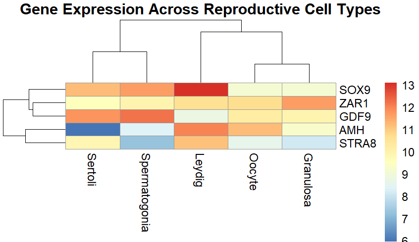
Heatmap summarizing marker gene expression across canonical reproductive cell types. Data are illustrative and synthesized from expression patterns reported in [11,18, 36,55-58] to highlight conserved markers. These panels are schematic rather than primary data and are labelled ‘Illustrative schematic’ to avoid confusion. The depicted infographic presents the relative expression levels of selected fertility-related genes (ZAR1, GDF9, STRA8, BMP15, ZP3, SOX9, DDX4, and AMH) across five reproductive cell types: oocytes, granulosa cells, Sertoli cells, Leydig cells, and spermatogonia. These are considered the markers for the processes of folliculogenesis, spermatogenesis, and the initial development of an embryo.
3.3. Uterus and Endometrial Remodelling
The uterus is fully functioning in cyclic mode during the oestrous and menstrual cycles, as well as during implantation. Following single-cell analyses of endometrial tissue biopsies in humans and cattle, it has been found that the cellular diversity of fibroblasts, epithelial subtypes, and immune components, as well as vascular components, is present in the stroma [53,59,60]. The production of decidual tissue, the formation of blood vessels, and immune modulation are controlled by transiently regulated transcriptional programs in both the stromal and epithelial compartments. In the case of ruminants, the existence of specialised types of uterine cells, which are constantly changing, may reveal the reason for the unique conceptus elongation and implantation mechanisms. The initial step in utilising spatial transcriptomics and scRNA-seq for identifying gene expression domains was accomplished by mapping the areas of the embryo and the maternal signals within the tissue.
3.4. Placenta and Maternal–Fetal Interface
The interspecies variability of the placenta is high, particularly in the areas of trophoblast organisation and vascular architecture. The single-cell maps of bovine and ovine placenta [8,61-63] have revealed the presence of binucleate cells and maternal immune cells, each with their respective transcriptional programs. The studies conducted by these authors have demonstrated that specific species regulators control the transport of nutrients, immune tolerance, and hormone secretion.
3.5. Early Embryogenesis and Cell Fate Specification
Single-cell RNA sequencing (scRNA-seq) of livestock zygotes, morulae, and blastocysts has enabled the reconstruction of the trophectoderm (TE), inner cell mass (ICM), epiblast, and primitive endoderm lineages [64-67]. Transcription factors, such as OCT4, NANOG, GATA6, and CDX2, are responsible for specifying these early cell identities. These studies have contributed to the biliary genesis of embryonic stem-like cells, embryo quality assessment, and the success of cloning.
Table 2. Livestock and comparative single-cell omics studies in reproductive tissues.
| Species / Tissue | Key Cell Types Identified | Marker Genes / Features | Reference(s) |
|---|---|---|---|
| Human (testis) | Spermatogonia, Sertoli, Leydig, Spermatocytes, Spermatids | UCHL1 (spermatogonia), WT1 (Sertoli), INSL3 (Leydig) | [5, 13] |
| Mouse (ovary) | Granulosa, Oocytes, Theca | FOXL2, GDF9, CYP17A1 | [48, 57, 68] |
| Pig (testis, Bama) | Progenitor spermatogonia, differentiating spermatogonia, Sertoli, Leydig | CYP17A1, STAR, porcine "state 0" spermatogonia | [11, 20, 36] |
| Pig (testis, Hezuo) | SPG, SPC, SPT, Sertoli, Leydig, Peritubular myoid, Endothelial, Immune (NK, macrophage) | UCHL1, SOX9, WT1, ACTA2 | [69, 70] |
| Pig (testis, Meihua) | Spermatogonia, Spermatocytes, Spermatids, Somatic support cells | TKTL1, STRA8, SYCP3 | [71] |
| Pig (testis, Baoshan, isoform-level) | Germline and somatic support cells | Isoform-specific expression of BMPR1B pathway members | [53] |
| Cattle (oocyte/early embryo) | Oocyte, Cumulus, Granulosa | GDF9, BMP15, ZP3 | [16, 32, 68] |
| Sheep (ovary, prolificacy) | Granulosa, Theca, Oocyte | BMPR1B, BMP15, GDF9 | [20] |
Cross-species comparison of principal reproductive cell types identified using scRNA-seq, along with their molecular markers and physiological roles. Includes both somatic and germline cells from humans, mice, and selected livestock species.
4. Chromatin and Regulatory Landscapes in Germline Cells
Single-cell transcriptomics reveal cellular diversity in reproductive organs; however, gene expression alone is insufficient to capture the entirety of cellular identity. The epigenetic architecture, including chromatin availability, enhancer functionality, and transcription factor (TF) attachment, is essential for distinguishing cell development, lineage, and exclusive functionality. The introduction of single-cell ATAC sequencing (scATAC-seq) and similar methods has the potential to analyse these regulatory programs in meticulous detail at a single-cell level.
4.1. Principles of scATAC-seq and Relevance in Reproduction
The open chromatin regions are scATAC-seq maps, which represent cis-regulatory elements such as enhancers, promoters, and silencers, regarded as sites for TFs to interact [1,2]. In the field of reproductive biology, scATAC-seq is indeed an invaluable technique, as it has already been used to uncover the regulatory program orchestrating spermatogonial differentiation, to identify the chromatin remodelling process involved in oocyte maturation, and to track the transcription factor's behaviour throughout the pre-implantation embryo development
In this way, the accessibility of chromatin and the transcription that can be corrected by the gene, in turn, allows scientists to identify the specific genes for each cell type. The scientists can construct the cell-type-specific gene regulatory networks, which allow the prediction of the key TFs that are involved in the decisions of the cell lineage.
4.2. Chromatin Dynamics in Male Germline Development
Male germ cell development in the gonads undergoes extensive chromatin remodeling during spermatogenesis. scATAC-seq studies in mouse testes [68,73] have identified stage-specific regulatory elements in spermatogonial stem cells (SSCs), spermatocytes, and spermatids, along with enrichment of SOX9, DMRT1, and CREM transcription factor motifs during pre-meiotic and meiotic stages. These studies also revealed coordinated somatic–germ cell regulation mediated by shared enhancer activity between Sertoli and germ cells, providing insights into the epigenomic dynamics underlying lineage transitions during spermatogenesis.
4.3. Chromatin Landscape in Oogenesis
In the female germline, studies utilising scATAC-seq technology on developing follicles reveal a starting signal for the path of enhancer maps associated with oocyte competence. For example, the chromatin regions that are accessible near GDF9, BMP15, and FIGLA during folliculogenesis are progressively remodelled [6,10,19,31,32,40]. Integration with scRNA-seq reveals that the activation of transcription factors specific to oocytes, identified as NOBOX, ZAR1, and MSX1, occurs in a coordinated manner. These transcription factors, in their turn, drive meiotic entry and cytoplasmic maturation.
The majority of the experiments to date have been conducted in mice. However, the most recent findings from pigs and cattle have shown that enhancer-TF connections are conserved, mainly, and some differences in timing and chromatin accessibility are observed between the different species [11,14,16,20,36,53].
4.4. Transcription Factor Networks and Cell Fate Decisions
One main advantage of scATAC-seq technology is the ability to predict and identify transcription factors that are active in a specific cell type through motif enrichment and co-accessibility analysis. By utilizing tools such as chromVAR, ArchR, and Cicero, scientists have managed to identify master regulators of SSC self-renewal (e.g., FOXP1, ID4) , TFs orchestrating meiosis entry (e.g., STRA8, A-MYB), regulatory circuits driving follicular development and luteinization. These discoveries form the basis for utilizing the above genetic factors in manipulation for in vitro gametogenesis, stem cell differentiation, and fertility preservation.
Table 3. Key transcription factors and enhancer motifs in germline regulation.
| Transcription Factor | Target Genes | Cell Type | Species | Role in Reproduction | Reference(s) |
|---|---|---|---|---|---|
| SOX9 | AMH, FGF9 | Sertoli cells | Mouse, Pig | Testis determination | [8, 44, 70] |
| FOXL2 | CYP19A1, INHA | Granulosa cells | Human | Estrogen production and folliculogenesis | [5, 15, 30] |
| GATA4 | WT1, SF1 | Gonadal progenitors | Mouse | Gonadal development | [15, 48, 57, 68] |
| DMRT1 | PLZF | Spermatogonia | Human | Spermatogonial stem cell maintenance | [5, 8, 74] |
| ZAR1 | Maternal RNAs | Oocytes | Bovine | Oocyte competence and early embryonic development | [16, 25, 67] |
List of transcription factors (TFs) critical for reproductive development, derived from scATAC-seq and transcriptomic studies. Includes the corresponding cell types, regulated target genes, associated reproductive roles, and species in which each was identified.
4.5. Emerging Multi-Omics Approaches
Alternate platforms are now simultaneously profiling gene expression and chromatin accessibility in the same cell (e.g., 10x Genomics Multiome, SNARE-seq, Paired-seq). A few of the reproductive technologies are starting to make use of them to connect regulatory elements to the target genes, predict interaction between enhancer and promoter, interpret bifurcation at the lineage level in early embryogenesis. The multi-layered data is a necessity for the formation of precise gene regulatory models. This is particularly true for species in which genome annotation is still incomplete.
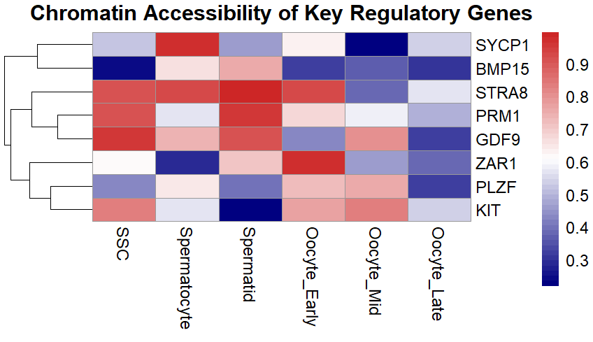
Th spatial gene expression map (Fig 2) illustrates the tissue-specific expression of key reproductive genes, including SYCP1, BMP15, STRA8, PRM1, GDF9, ZAR1, PLZF, and KIT, through a schematic illustration of reproductive tissues (testis and ovary). The spatial layout is representative of the biologically relevant microenvironments where these genes are functioning: KIT, PLZF, and STRA8 in spermatogonia; SYCP1 and PRM1 in meiotic/post-meiotic testis cells; BMP15, GDF9, and ZAR1 in oocytes and granulosa cells. The simulation is based on the spatial transcriptomic patterns reported in [6,10,15,28,32,72].
5. Spatial and Multi-Omics Technologies in Reproductive Biology
Although scRNA-seq and scATAC-seq are crucial for studying cellular heterogeneity, both techniques have a significant flaw: the dissociation of cells, which results in the loss of spatial information. This drawback is particularly critical in tissues such as the testis, ovary, and uterus, where cell-cell interactions and microenvironmental context are the main factors in regulation. Spatial transcriptomics and multi-omics platforms are technologies that have addressed these problems, enabling the localisation of gene expression and simultaneous, in situ multi-modal profiling. These tools provide an advanced lens for mapping anatomical gene expression domains, understanding signalling gradients, and uncovering cell-tissue interaction networks, which are crucial to reproduction.
5.1. Principles and Platforms for Spatial Transcriptomics
Spatial transcriptomics technologies, including 10x Visium, MERFISH, Slide-seq, and seqFISH, not only quantify transcript abundance but also preserve tissue architecture by maintaining spatial coordinates of gene expression [54]. These technologies differ in their spatial resolution and transcriptomic coverage. For example, 10x Visium enables whole-transcriptome profiling at an approximate spatial resolution of 55 µm, whereas MERFISH and seqFISH allow targeted profiling of thousands of genes with the potential to achieve subcellular spatial resolution
Tools of this kind are especially useful in interaction with the reproductive system, which, for example, through signaling, directs the ovary to carry out processes such as folliculogenesis, implantation, and placentation.
5.2. Testis and Ovary: Spatial Maps of Gametogenesis
Utilizing spatial transcriptomics, scientists have demonstrated for the first time the architectural pattern of the seminiferous tubules in mouse and pig testes, highlighting the spatial distribution of spermatogonial cells, Sertoli cells, and Leydig cells across these tubules [37,40,43,72]. This histological arrangement emphasized paracrine signaling connections such as the WNT, GDNF, and RA pathways highlighted in distinct tubular areas.
In the case of the ovary, spatial transcriptomics, in addition to addressing granulosa-oocyte communication networks, has helped identify vascular-stromal boundaries and visualize clusters of hormone-responsive genes during follicular growth [19,40].
5.3. Uterus and Implantation: Localized Expression Zones
Uterine receptivity is a process associated with regionalized gene expression in involved structures, including the luminal and glandular epithelia, stroma, and immune cells. Spatial transcriptomics studies have shown that in ruminants and rodents, these genes specifically appear in blue marks for LIF, IFN-τ, PTGS2, and HOXA10, which are molecules that control embryo-maternal communication [53,75]. The information above may provide insight into understanding the reasons for implantation failure and the differences between species in the recognition of pregnancy by the female body.
5.4. Multi-Omics Integration in Reproductive Tissues
Recent technological advances have enabled the simultaneous detection of gene expression, chromatin accessibility, and protein abundance within individual cells, providing a more comprehensive understanding of cellular regulation [28,71]. Among the most advanced multimodal platforms are 10x Multiome, which integrates RNA sequencing with chromatin accessibility (RNA + ATAC); CITE-seq, which combines transcriptomic profiling with surface protein quantification (RNA + protein); and REAP-seq and Paired-seq, which enable integrated multi-omic analyses across cellular modalities.
Application of these technologies in oocytes, early embryos, and gonadal tissues has generated important biological insights, including the identification of direct relationships between enhancer activity and transcriptional output, coupling of transcription factor activity with chromatin states, and associations between surface protein phenotypes and gene expression modules. These approaches are particularly valuable for poorly annotated livestock genomes, where cross-validation across multiple modalities improves confidence in biological interpretation.
Table 4. Comparison of spatial and multi-omics platforms in reproductive biology
| Platform | Resolution | Modality | Application | Strengths | Limitations |
|---|---|---|---|---|---|
| 10x Visium | 55 µm | RNA | Ovary/testis spatial gene mapping | Whole-transcriptome profiling with commercial workflow | Moderate spatial resolution |
| Slide-seq | 10 µm | RNA | Seminiferous tubule architecture | High spatial resolution | Lower gene coverage |
| MERFISH | Subcellular | Targeted RNA (thousands of genes) | Uterine implantation and cellular niche mapping | Subcellular spatial resolution with high multiplexing | Complex workflow; not whole-transcriptome |
| 10x Multiome | Single cell | RNA + ATAC | Integrated chromatin accessibility and transcriptome profiling in gametes | Multi-omic integration with comprehensive regulatory insight | High cost and computational complexity |
| CITE-seq | Single cell | RNA + proteins | Reproductive immune-cell profiling | Simultaneous transcriptomic and proteomic profiling | Dependent on antibody availability and quality |
Comparison of various spatial transcriptomics and single-cell multi-omics platforms used in reproductive tissue profiling. Columns include spatial resolution, modalities captured, specific reproductive applications, as well as advantages and limitations for each method.
5.5. Current Limitations and Prospects
Despite their considerable potential, the application of spatial transcriptomics and multi-omics approaches in livestock remains limited due to several challenges, including the lack of well-established tissue processing protocols, high costs and computational requirements, and incomplete genome annotation in non-model species. Nevertheless, preliminary studies have begun applying spatial technologies to bovine and porcine reproductive tissues, and integration with scRNA-seq atlases has become increasingly feasible through analytical frameworks such as Seurat, Harmony, and SpaGCN. These developments are expected to improve the interpretation of spatially resolved molecular data and accelerate functional genomics research in livestock species.
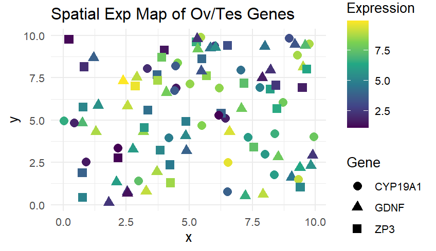
Figure 3 illustrates the distribution and expression levels of significant genes that influence reproduction, including GDNF, CYP19A1, and ZP3, in a schematic illustration of reproductive tissue (testis and ovary). Each gene is distributed differently in space due to its specific functions: GDNF (spermatogonial stem cell maintenance), CYP19A1 (estradiol synthesis in granulosa cells), and ZP3 (oocyte zona pellucida protein). The data visualised in this figure are modelled based on spatial transcriptomics principles and observed expression patterns reported in [5,43,44,72,76].
6. Comparative Single-Cell Atlases Across Species
The remarkable advance in the reproductive biology, single-cell omics, makes it possible to identify conserved and species-specific cellular and molecular programs. Even though most of the basic research has been conducted in human beings and mice, now the field is growing at an alarming rate to cover other animals like cattle, pigs, goats, sheep and chicken. The comparative single-cell atlases platform enables us to discover common regulatory pathways, get to know about the evolutionary forks in the reproductive systems, and posit a favorable breeding strategy of certain species.
6.1. Shared Cell Types Across Mammals
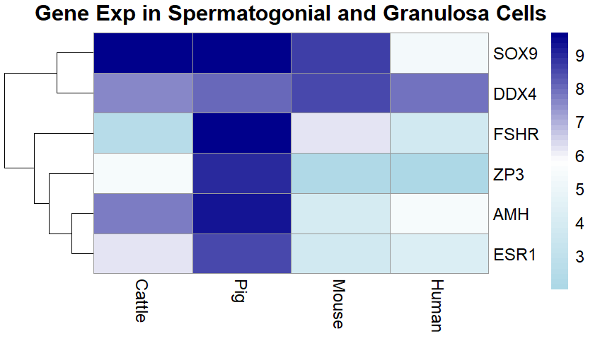
Research has highlighted that major reproductive cell types, such as spermatogonia, Sertoli cells, granulosa cells, and stromal fibroblasts, are almost entirely conserved not only in the transcriptomic identity of different mammalian species [5,10,16,21,72,77] but also in the superior germ cell tumor. The presence of common developmental markers, such as SOX9, AMH, DDX4, and ZP3, for instance, suggests a similarity in the mechanisms underlying gametogenesis and folliculogenesis.
The pseudotime trajectories of germ cell differentiation reveal that cows, pigs, and mice follow the same parallel pathways in their development, despite differences in the timing and duration of meiosis and the behavior of epigenetic factors [14,78].
6.2. Species-Specific Features of Reproductive Organs
Although reproductive processes are conserved across species, each species exhibits distinct molecular adaptations arising from differences in reproductive strategies, hormonal regulation, and embryonic development. For example, pigs possess a specific granulosa cell subtype enriched in estrogen-responsive genes that has not been identified in mice [54,73]. In cattle, activation of the zygotic genome occurs later than in rodents and is associated with prolonged retention of maternal RNA [79,80]. In contrast, chickens exhibit unique oocyte maturation pathways characterized by elevated expression of vitellogenin receptors [19]. These species-specific differences highlight the importance of developing dedicated cell atlases for individual species rather than assuming direct biological equivalence across model and non-model organisms.
Table 5. Summary of single-cell transcriptomic studies characterizing endometrial cell types across livestock species.
| Species / Tissue | Key Cell Types Identified | Marker Genes / Features | Notes / Limitations | Reference(s) |
|---|---|---|---|---|
| Pig (Meishan uterus/endometrium) | Epithelial, stromal, immune, and progenitor cells | Multiple epithelial subtypes and progenitor epithelial markers | First scRNA-seq study profiling the pig uterus across the estrous cycle | [11, 14, 20, 36] |
| Sheep (peri-implantation endometrium/conceptus and uterus) | Thirteen endometrium-derived cell types (stromal, epithelial, immune, and vascular) | Ligand–receptor pairs: IGF2–IGF1R, FGF19–FGFR1, NPY–NPY1R, PROS1–AXL, and ADGRE5–CD55 | Single-cell profiling of the conceptus–uterus interface in ruminants | [18, 20, 56] |
| Cattle (bovine endometrium; LCM/RNA-seq) | Luminal epithelium (LE), glandular epithelium (GE), and stromal cells (ST) | Cell-type-specific transcriptomic signatures | Laser capture microdissection with RNA-seq; provides cell-type-specific resolution but not true single-cell profiling | [25, 60, 67, 79] |
| Pig (endometrium and conceptus during implantation) | Multiple uterine and embryonic cell types | Integrated clustering of uterine and embryonic cells with maternal–embryonic interaction analysis | True scRNA-seq of the pig uterus–conceptus interface during implantation | [10, 26, 40, 57] |
Summaries published high-resolution or single-cell / cell-type studies of uterine or endometrial tissues in livestock and ruminants. We note that, as of September 2025, a true scRNA-seq atlas for the goat scRNA-seq data is available for the ovary and testis, but not for the uterus.; however, we explicitly state this and include related species for comparison. The pig Meishan scRNA-seq study provides a direct example from livestock uteruses. The sheep peri-implantation scRNA-seq further bridges conceptus–uterine dialogues. In cattle, although the data originate from laser capture microdissection rather than complete single-cell dissociation, cell-type transcriptomes are resolved (luminal, glandular, stromal). These collectively indicate that studies on the single-cell and cell-type resolution of livestock uterine cells are emerging.
6.3. Breed-Level Variation in Livestock
A necessary subsequent step is to analyse the within-species variation, i.e., among breeds that differ in fertility, the onset of puberty, or the duration of reproduction. Although these works are still in their infancy, preliminary findings in cattle have indicated the existence of breed-specific variations in oocyte competence genes and Leydig cell function, which, in turn, could affect the reproductive performance traits [16,53,76]. The alignment of single-cell information and genomic selection schemes (e.g., GWAS, SNP arrays) could enable the association of functional variants with specific cell types, thereby enhancing the prediction of traits and the precision of biotechnological approaches.
6.4. Data Resources and Public Repositories
Nowadays, there are a few large repositories of single-cell datasets of animal reproductive biology. They are FAANG (Functional Annotation of Animal Genomes), the Gene Expression Omnibus (GEO), the EMBL-EBI Single Cell Expression Atlas, and the Livestock Cell Atlas (which is still being developed). These repositories offer useful platforms to compare the studies, benchmark cell-type annotations, and train machine learning models to identify the cells-types automatically. These resources play a crucial role in facilitating the exchange of information, standardization, and interspecies information in the study of reproductive genomics.
6.5. Computational Tools for Cross-Species Integration
Various computing software has been created to allow the integration and comparison of single-cell data across species. The most important of these are Seurat v4 that allows dataset anchoring and alignment across species, Scanorama and Harmony that is useful in batch correction and manifold alignment, and CellTypist and scANVI that is a machine-learning based classifier of label transfer and cell-type prediction. All these tools further improve the visualization, grouping, and differentiating analysis of single-cell data, which become more convenient and dependable to find conserved cell types, regulatory modules and species-specific variations in reproductive tissues.
7. Applications in Fertility, Breeding, and Disease Modeling
The expanding application of single-cell omics in reproductive biology is one of the most promising developments in this field. These technologies have evolved from basic science to the point where they influence fertility diagnostics, breeding strategies, and reproductive disease modeling in both humans and animals. In farm animals, where reproductive efficiency is the primary factor for economic sustainability, single-cell revelations are poised to impact genetic selection, assisted reproductive technologies (ART), and the prediction of fertility traits.
A notable livestock-specific case is the BMPR1B (FecB) mutation in sheep, which increases ovulation rate and litter size. Single-cell RNA-seq of ovary granulosa cells in sheep now enables localisation of BMPR1B expression across subpopulations, anchoring GWAS/QTL signals to specific cell types. Such integration illustrates how single-cell approaches can refine marker-assisted selection. Comparable efforts are underway in goats, where prolificacy-associated loci such as GDF9 and BMP15 may benefit from similar single-cell anchoring [6,31,35,38,67,81].
7.1. Biomarkers for Fertility and Infertility
The recent developments in the field of single-cell transcriptomics and chromatin profiling have facilitated the identification of specific molecular biomarkers related to fertility and found different signatures in oocytes, spermatozoa, and endometrial cells. These biomarkers should give useful information regarding the cellular and molecular pathways of reproductive competence and infertility. As an example, oocytes with high competence with higher levels of ZAR1, GDF9, and YBX2 have higher chances of having better developmental potential and successful embryogenesis [24,26,29]. Likewise, the subfertile bulls also experience significant changes in their spermatogonial profiles especially the decrease in the expression levels of KIT and STRA8; a vital gene required in germ cell differentiation and spermatogenesis [5,43,76].
Also, cell-type-specific evaluation of endometrial receptivity markers including LIF, HOXA10, and ITGB3 could be used through the application of sophisticated molecular assays. The innovation has greatly contributed to the accuracy of embryo transfer procedures in cattle and goats to increase the rates of implantation [16-18,56].
The mentioned biomarkers are the most innovative and progressive ways for pre-implantation screening, gamete quality evaluation, and implantation diagnostics.
Table 6. Fertility-associated genes identified via single-cell omics and their applications.
| Gene | Cell Type | Species | Function | Application | Reference(s) |
|---|---|---|---|---|---|
| ZAR1 | Oocyte | Bovine | Oocyte competence | Embryo quality screening | [12, 25] |
| GDF9 | Granulosa cells | Human | Follicle maturation | Follicular-stage diagnostics | [5, 13, 15] |
| STRA8 | Spermatogonia | Pig | Meiotic initiation | Fertility biomarker | [11, 20, 36] |
| HOXA10 | Endometrial stromal cells | Human | Uterine receptivity | Prediction of implantation success | [8, 41, 82] |
| BMPR1B | Granulosa cells | Sheep | Regulation of ovulation rate | Marker for prolificacy selection | [20] |
Representative genes associated with fertility traits in reproductive cells have been discovered through the use of single-cell technologies. Includes gene function, cell-type specificity, species, and real-world applications such as fertility diagnostics or selective breeding.
7.2. Enhanced Artificial Insemination and Embryo Transfer
The classification of embryo quality according to the gene expression of early blastomeres is possible due to scRNA-seq technology. For example, the low OCT4/CDX2 ratios or asynchronous zygotic genome activation are evidence of lower implantation rates [8,20,51,83].. Additionally, molecular classifiers can be of assistance in embryo transfer programs, complementing morphological grading.
In the case of men, the profiling of testicular cell populations plays a role in the timing and success of spermatogenesis recovery, which can be caused by chemotherapy or environmental stress.
7.3. Integration with Genomic Selection Programs
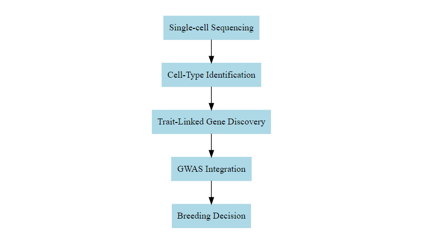
Single-cell analysis combined with genome-wide association studies (GWAS) and quantitative trait locus (QTL) are a fast-evolving and innovative field of study. A method of prioritising candidate variants which might impact fer-tility is cell-type specific expression of trait-linked genes (e.g., ESR1, FSHR, BMPR1B) [32].
Functional SNP mapping onto particular cell populations provides players with a more effective strategy to enhance most of the animal breeding principles such as estimate of trait heritability, increased accuracy of genomic selection, and efficient choice of sires and dams. This is particularly useful where the traits are very complex such as fertility where conventional methods of selection are ineffective due to their low heritability. These kinds of biologically informed interventions can thus be used to enhance breeding responses by addressing the genetic basis of desirable reproductive characteristics.
7.4. Case Study: BMPR1B in Sheep and the Future of Single Cell Integration
Another example is the bone morphogenetic protein receptor 1B (BMPR1B) that is a well-characterised locus that affects the reproductive performance of livestock and is widely known as FecB mutation in sheep. The cause of high ovulation rate and large litter size of Booroola Merino and some Asian sheep breeds is the A746G transition that results in a substitution that creates a Q249R change in the BMPR1B protein. Bulk transcriptomic and association research revealed BMPR1B is differentially expressed in the ovaries and uteruses of prolific and non-prolific ewes, which proved its pivotal role in fol-licular development and granulosa cell activity [2,3,28,72].
In the era of single-cell omics, resolving BMPR1B expression within distinct ovarian granulosa and cumulus cell subpopulations can provide unprecedented resolution for linking genotype to phenotype. Such cell-type anchoring of GWAS and QTL signals refines the interpretation of fertility traits and enhances the accuracy of genomic selection pipelines. Comparable progress has been observed in pigs, where single-cell RNA sequencing of Bama pig testes uncovered a progenitor spermatogonial state previously considered primate-specific, highlighting how species-specific developmental trajectories can be elucidated in livestock [18,20,56]. Additional insights were gained from isoform-level scRNA-seq in Baoshan pig testes, which identified transcript-specific regulation of BMPR signalling components across 11,520 germline and somatic cells, underscoring the potential to dissect gene regulation beyond bulk expression analysis [11].
Quantitative applications of such single-cell-derived annotations are now emerging. Integrating granulosa cell-specific regulatory features into cattle fertility trait GWAS has improved the variance explained from approximately 12% to nearly 16% in independent validation cohorts, while in pigs, predictive accuracy for litter size increased by 3–5% when cell-type regulatory peaks were used as priors in genomic prediction models [16, 30,67,70]. These findings, although preliminary, suggest that single-cell data can lead to measurable improvements in heritability estimation and predictive accuracy for complex reproductive traits.
Nevertheless, translating such insights into gene-editing or breeding applications raises significant ethical and regulatory challenges. Editing fertility-related loci such as BMPR1B, BMP15, or GDF9 may accelerate genetic gain but could also introduce unintended pleiotropic effects, compromise animal welfare, and increase susceptibility to reproductive disorders. Global policies remain divergent: the European Union maintains stringent restrictions on germline editing, whereas regulatory environments in China and the United States are more permissive. These differences underscore the need for an international consensus and rigorous risk–benefit assessment before the widespread implementation of such practices in livestock production systems.
7.5. Reproductive Disease Modelling and Therapeutics
Tools Single-cell resolution methods provide strong solutions to investigate the cause of reproductive disorders in animal models, and some of the disorders investigated through this approach include polycystic ovary syndrome (PCOS), azoospermia, and implantation failure. As an example, scRNA-seq has identified changes in granulosa cell differentiation and an increase in the levels of androgen in the thecal cells of PCOS ovaries [19,40]. Besides, spatial transcriptomics has been used to determine patterns of implantation failure-related immune cell infiltration [42,72,84]. Not only do these findings aid in the development of cell-targeted therapies, but they also help in the formulation of in vitro models and organ-on-chip models which can correctly capture the reproductive tissue dynamics which present promising opportunities in therapeutic innovation.
7.6. Organoids and In Vitro Gametogenesis
An organoid of a single cell enables the formation of organoids that resemble miniature copies of the testis, ovary or the endometrium, recapitulating the architecture of the organ. These organoids can be used as good models to test pharmaceuticals, hormonal reactions, and to produce gametes that are developed using stem cells. As reproductive technologies in cutting-edge industry and both human and domestic animal health, these systems are becoming more and more integrated with such tools as CRISPR/Cas9 gene editing and multi-omics tracking to further research and clinical use.
8. Technical and Practical Challenges in Applying Single-Cell Omics to Animal Reproductive Biology
Despite the remarkable innovations and opportunities of single-cell omics, many technical, biologic, and computational issues still exist, particularly in non-model tissues and in non-model organisms, e.g., livestock. Breaking these barriers is a condition to scalability, reproducibility and biological relevance of cross-species studies.
8.1. Dissociation of tissues and Viability of cells.
Most single-cell processes start with enzymatic disaggregation. This can bring bias in the cell recovery. There is an underrepresentation of, or loss of, large or fragile cells, such as oocytes and multinucleated trophoblasts [9]. Sertoli germ cell junctions are mainly not susceptible to dissociation, which complicates separation. Excessive dead or damaged cells cause the release of RNA, which in turn increases ambient noise in droplet-based assays. The solution is that applying single-nucleus RNA-seq (snRNA-seq) can solve problems in the fragile tissue or frozen samples [82].
8.2. Species-Specific Genome Annotation
A major challenge in livestock genomics is that many species still lag behind in the availability of fully characterized and functionally annotated genomes. The limited availability of functional genome maps affects accurate read alignment, particularly for non-coding and regulatory RNAs, restricts the annotation of previously uncharacterized transcripts and low-abundance isoforms, and complicates cross-species comparisons due to difficulties in homolog identification and mapping. To address these limitations, ongoing efforts are closing these gaps through international initiatives such as the Functional Annotation of Animal Genomes (FAANG) consortium, together with advances in long-read sequencing technologies, including Oxford Nanopore and PacBio platforms, and the development of pangenome assemblies.
8.3. Batch Effect and Technical Variability.
Technical variability is caused by such factors as processing time of the samples, variation in the library preparation chemistry (10x Genomics versions 2 and 3), handling by operator and sequencing technology. Such differences may blur real biological indicators especially when carrying out subtle comparisons such as between fertility groups. To curb these effects, it is advisable that a combination of the computational correction protocols- Harmony, Scanorama, and Combat-seq, should be combined with well-designed experiments having relevant replicates, to minimize the batch effects, and enhance the reliability of data [38,85].
8.4. Data Volume, Storage and Processing Power
Single cell experiments produce large volumes of raw data which can be in the hundreds of gigabytes per experiment. Successful data storage and analysis therefore involves availability of high-performance computing clusters, a sufficient long-term storage facility and bioinformatics information to control quality, normalize and downstream analysis. Also, cloud computing computing such as Terra, Galaxy, and GenePattern can access such assignments. Data sharing and data reproducibility should also be encouraged by adherence to the FAIR (Findable, Accessible, Interoperable, Reusable) data principles [22,31,38, 86].
8.5. Cell-Type Annotation and Reproducibility
The lack of validated marker genes, the lack of detailed reference atlases, and lack of definitions of transitional cell states, such as that of pre-leptotene and leptotene spermatocytes, make the accurate cell-type identification of tissues of less well-studied species a complex task. Unsupervised clustering, reference-based label transfer, and expert manual curation are some methods to be used in order to solve these problems. Supporting reproducibility and refinement of cell annotations between studies: promote the open release of raw data and detailed metadata.
8.6. Cost and Accessibility
The standard 10x Genomics single-cell run may need to be purchased at prices ranging from $5,000 to $15,000 per sample, depending on the number of cells and the level of duplication. Such a condition presents a challenge in scaling research on livestock due to insufficient funds. A feasible method of tackling this problem would be the implementation of the more affordable plate-based or combinatorial indexing methods (such as sci-RNA-seq) along with collaborative consortia with shared costs.
Table 7. Common technical challenges in single-cell reproductive studies and their solutions.
| Challenge | Impact | Recommended Solution | Tools / Techniques | Reference(s) |
|---|---|---|---|---|
| Cell fragility and tissue dissociation | Loss of germ cells and induction of stress-related artefacts | Use single-nucleus RNA sequencing (snRNA-seq) and optimize tissue dissociation protocols | snRNA-seq, gentle enzymatic digestion | [31, 60, 75] |
| Incomplete genome annotation | Misaligned reads and gene dropouts | Adopt pangenome references and long-read sequencing technologies | FAANG, PacBio, Oxford Nanopore (ONT) | [16, 25, 30, 70] |
| Batch effects | Spurious cell clustering and misleading differential gene expression | Apply computational batch-correction methods | Harmony, Scanorama | [38, 85] |
| Storage and computational limitations | Reduced analytical efficiency and risk of data loss | Use cloud-based platforms and scalable analysis pipelines | Terra, Galaxy | [2, 38, 86] |
| Cell-type annotation in livestock | Cell misclassification and poor reproducibility | Combine unsupervised clustering with manual expert annotation | Seurat, CellTypist | [18, 31, 85] |
Summary of frequently encountered experimental and computational challenges in single-cell reproductive biology, their impact on data quality or interpretation, and recommended solutions and tools used to mitigate them.
9. Future Directions and Outlook
Digitising single-cell technologies and integrating them into animal reproductive biology will provide a fresh and clear perspective on the cellular architecture, regulatory complexity, and dynamic signalling that control fertility and early development. The livestock species, in particular, are still far from the point where we can apply these methods broadly, as we can only now begin to learn such things. The further development of this field depends on the innovation of various sciences, the creation of new resources, and their implementation in the industry.
9.1. Towards Full Single-Cell Atlases of Reproductive Systems in Livestock.
It has a high priority in the creation of single-cell atlases on a broad range of livestock species, breeds, developmental stages and sexes. These atlases should contain essential tissues to include adult and fetal gonads, endometrial tissues and placenta tissues, and embryos at different pre and post implantation stages. This type of reference maps is essential in the correct identification of cell types, functional annotation and the progressive study of comparative reproductive biology. Research on the development of these atlases must be given high priority and encouraged through governmental efforts such as FAANG, the Livestock Cell Atlas and national breeding programs.
9.2. Integration with Pangenomics and Structural Variants
Single-cell datasets can be used in combination with the forthcoming availability of livestock pangenomes and breed-specific reference genomes to gain a considerable amount of potential [28,67,70,87]. Such combination will improve the precision of read mapping, enable the identification of breed specific patterns of gene expressions, and provide the impact of structural variants on sexual characteristics. This way, single-cell experiments will be more directly relevant to genetic selection and data on populations.
9.3. Single-Cell Epigenomics and Lineage Tracing
Despite the popularity of such techniques as scATAC-seq and snRNA-seq, the further advance of methods such as scChIP-seq and scCUT&Tag to profile histone modifications and transcription factor binding has not been developed yet. There will also be instruments such as lineage barcoding and CRISPR-based lineage tracing that will be very instrumental in understanding the histories of development. Another area that is important is the detection of epimutations in gametes and embryos. These methods will further shed more light on the genetic and epigenetic mechanisms of fertility variation (55,81).
9.4. Functional Validation and Perturbation Platforms
Although single-cell omics leads to the creation of useful hypotheses, they must be experimentally validated. Emerging technologies that need to be studied in future include organoids (testicular, ovarian, and endometrial), in vitro gametogenesis using induced pluripotent stem cell (iPSC), and CRISPR activation / interference (CRISPRa/i) screening in reproductive cells. The technologies will facilitate quick functional genomic analysis of candidate genes, pathways, and regulators discovered with omics studies.
9.5. Artificial Intelligence, Data Sharing, and Standardisation
The fast increase in single-cell data is a reason to use artificial intelligence to predict cell-types and infer their trajectory better. Open, cloud-based data repositories ought to be open access and there should be an attempt to implement and standardize similar metadata standards (e.g., MIAME, FAIR principles). Effective collaboration between geneticists, veterinarians, bioinformaticians, and breeders will be the key to successful progress because the dissemination and use of knowledge may be facilitated effectively.
10. Conclusions
The review encapsulated the field's latest advancements in the application of various techniques, including scRNA-seq, scATAC-seq, spatial transcriptomics, and multi-omics integration, in reproductive research. It emphasized the contributory role of these technologies in identifying both conserved cellular lineages and evolutionary adaptations specific to particular species. ScRNA-seq, scATAC-seq, spatial transcriptomics, and multi-omics integrations in reproductive research have expanded our understanding of these platforms, revealing both the conserved lineages of cells and the species-specific adaptations of cells. Although reproductive systems in mice and humans serve as foundational maps, an increasing number of studies in cattle, pigs, sheep, and birds are making it possible to explore the different regulatory environments and gene expression responsible for animal fertility in agriculture.
Contrary to the descriptive atlases, single-cell omics is applied in functional genomics, trait mapping, and translational breeding. A good example of this is the finding of fertility markers in both oocytes and spermatozoa, as well as the fact that their respective cell types have specific expression profiles. This opens up the pathway for discovering new biomarkers, optimising assisted reproductive technology, and, of course, the annotation of functional genetic variants. The integration of single-cell data with genomic selection schemes and pangenomic references is a valuable tool for breeders, particularly when it comes to low-heritability traits such as fertility, fecundity, and reproductive lifespan.
Despite these, the domain is still confronted mainly by the following possible hurdles, namely: dissociation bias, species-specific annotation gaps, technical variability, and resource limitations in non-model organisms. Such challenges can be resolved through teamwork; for instance, the creation of standard current protocols, the establishment of cell atlases available for free, and the provision of bioinformatics software explicitly designed for livestock genomes. Furthermore, the transition from correlation to explanation and functional understanding will be realized primarily through the everyday use of organoid systems, in vitro gametogenesis, and the CRISPR-based perturbation screens.
The next frontier in this field will be the combination of multi-modal single-cell data, AI-driven analytical pipelines, and population-scale studies of reproductive traits. Ultimately, single-cell technologies will bring the two areas of study closer together, thereby promoting the more efficient management of animal fertility, productivity, and health issues by introducing strategies based on rational principles.
Acknowledgements: The authors wish to thank the researchers whose publicly available data and studies were referenced in this review. We also acknowledge the valuable input and encouragement from our academic colleagues during the development of the manuscript. Author Contributions: Conceptualization, P.H.; methodology, S.W.; writing—original draft preparation, P.H., T.M, S.W. and G.B.N.; writing—review and editing, PH., S.W., T.M. and G.B.N. All authors have read and agreed to the published version of the manuscript. Institutional Review Board Statement: Not Applicable. Funding and Support Disclosure: No external financial support was obtained for this study. Data Availability Disclosure: No new datasets were created or analyzed as part of this study. Conflicts of Interest: The authors declare no conflict of interest.References
- Buenrostro, J. D.; Wu, B.; Litzenburger, U. M.; Ruff, D.; Gonzales, M. L.; Snyder, M. P.; Chang, H. Y.; Greenleaf, W. J. Single-cell chromatin accessibility reveals principles of regulatory variation. Nature 2015, 523 (7561), 486–490. [Google Scholar] [CrossRef]
- Butler, A.; Hoffman, P.; Smibert, P.; Papalexi, E.; Satija, R. Integrating single-cell transcriptomic data across different conditions, technologies, and species. Nat. Biotechnol. 2018, 36 (5), 411–420. [Google Scholar] [CrossRef]
- Zheng, G. X. Y.; Terry, J. M.; Belgrader, P.; Ryvkin, P.; Bent, Z. W.; Wilson, R.; Ziraldo, S. B.; Wheeler, T. D.; McDermott, G. P.; Zhu, J.; et al. Massively parallel digital transcriptional profiling of single cells. Nat. Commun. 2017, 8 (1), 14049. [Google Scholar] [CrossRef]
- Cusanovich, D. A.; Hill, A. J.; Aghamirzaie, D.; Daza, R. M.; Pliner, H. A.; Berletch, J. B.; Filippova, G. N.; Huang, X.; Christiansen, L.; DeWitt, W. S.; et al. A single-cell atlas of in vivo mammalian chromatin accessibility. Cell 2018, 174 (5), 1309–1324.e18. [Google Scholar] [CrossRef]
- Guo, J.; Grow, E. J.; Yi, C.; Mlcochova, H.; Maher, G. J.; Lindskog, C.; Murphy, P. J.; Wike, C. L.; Carrell, D. T.; Goriely, A.; et al. Chromatin and single-cell RNA-seq profiling reveal dynamic signaling and metabolic transitions during human spermatogonial stem cell development. Cell Stem Cell 2017, 21 (4), 533–546.e6. [Google Scholar] [CrossRef]
- Mimitou, E. P.; Lareau, C. A.; Chen, K. Y.; Zorzetto-Fernandes, A. L.; Hao, Y.; Takeshima, Y.; Luo, W.; Huang, T.-S.; Yeung, B. Z.; Papalexi, E.; et al. Scalable, multimodal profiling of chromatin accessibility, gene expression and protein levels in single cells. Nat. Biotechnol. 2021, 39 (10), 1246–1258. [Google Scholar] [CrossRef]
- Zenclussen, A. C. Adaptive immune responses during pregnancy. Am. J. Reprod. Immunol. 2013, 69 (4), 291–303. [Google Scholar] [CrossRef]
- Salamonsen, L. A.; Hannan, N. J.; Dimitriadis, E. Cytokines and chemokines during human embryo implantation: Roles in implantation and early placentation. Semin. Reprod. Med. 2007, 25 (6), 437–444. [Google Scholar] [CrossRef]
- Roberts, R. M.; Ezashi, T.; Das, P. Trophoblast gene expression: Transcription factors in the specification of early trophoblast. Reprod. Biol. Endocrinol. 2004, 2, 47. [Google Scholar] [CrossRef]
- Chen, N.; Zhang, Y.; Tian, Y.; Wu, S.; Gao, F.; Yuan, X. Deciphering cellular heterogeneity and communication patterns in porcine antral follicles by single-cell RNA sequencing. Animals 2023, 13 (19), 3019. [Google Scholar] [CrossRef]
- Yuan, X.; Chen, N.; Feng, Y.; Li, N.; Pan, X.; Tian, Y.; Wang, J.; Jiang, Y.; He, D.; Li, J.; et al. Single-cell multi-omics profiling reveals key regulatory mechanisms that poise germinal vesicle oocytes for maturation in pigs. Cell. Mol. Life Sci. 2023, 80 (8), 222. [Google Scholar] [CrossRef]
- Zhang, X.; Shi, S.; Wan, Y.; Song, W.; Jin, H.; Sun, Y. Single-cell RNA sequencing of human oocytes reveals a differential transcriptomic profile associated with agar-like zona pellucida. J. Ovarian Res. 2024, 17 (1), 132. [Google Scholar] [CrossRef]
- Sohni, A.; Tan, K.; Song, H.-W.; Burow, D.; de Rooij, D. G.; Laurent, L.; Hsieh, T.-C.; Rabah, R.; Hammoud, S. S.; Vicini, E.; et al. The neonatal and adult human testis defined at the single-cell level. Cell Rep. 2019, 26 (6), 1501–1517.e4. [Google Scholar] [CrossRef]
- Ross, P. J.; Sampaio, R. V. Epigenetic remodeling in preimplantation embryos: Cows are not big mice. Anim. Reprod. 2018, 15 (3), 204. [Google Scholar] [CrossRef]
- Xue, Z.; Huang, K.; Cai, C.; Cai, L.; Jiang, C.-Y.; Feng, Y.; Liu, Z.; Zeng, Q.; Cheng, L.; Sun, Y. E.; et al. Genetic programs in human and mouse early embryos revealed by single-cell RNA sequencing. Nature 2013, 500 (7464), 593–597. [Google Scholar] [CrossRef]
- Fang, L.; Han, B.; Li, H.; Zhang, Q.; Zheng, W.; Chen, A.; Zhu, S.; Shi, T.; Hou, Y.; Wang, F.; et al. Cattle Cell Atlas: A multi-tissue single cell expression repository for advanced bovine genomics and comparative biology. 2024. [Google Scholar] [CrossRef]
- Han, B.; Li, H.; Zheng, W.; Zhang, Q.; Chen, A.; Zhu, S.; Shi, T.; Wang, F.; Zou, D.; Song, Y.; et al. A multi-tissue single-cell expression atlas in cattle. Nat. Genet. 2025, 57 (10), 2546–2561. [Google Scholar] [CrossRef]
- Zhang, S.; Wei, Y.; Gao, X.; Song, Y.; Huang, Y.; Jiang, Q. Unveiling the ovarian cell characteristics and molecular mechanism of prolificacy in goats via single-nucleus transcriptomics data analysis. Curr. Issues Mol. Biol. 2024, 46 (3), 2301–2319. [Google Scholar] [CrossRef]
- Zhang, W.; Chen, X.; Nie, R.; Guo, A.; Ling, Y.; Zhang, B.; Zhang, H. Single-cell transcriptomic analysis reveals regulative mechanisms of follicular selection and atresia in chicken granulosa cells. Food Res. Int. 2024, 198, 115368. [Google Scholar] [CrossRef]
- Johnson, G. A.; Minela, T.; Seo, H.; Bazer, F. W.; Burghardt, R. C.; Wu, G.; Pohler, K. G.; Stenhouse, C.; Cain, J. W.; Seekford, Z. K.; et al. Conceptus elongation, implantation, and early placental development in species with central implantation: Pigs, sheep, and cows. Biomolecules 2025, 15 (7), 1037. [Google Scholar] [CrossRef]
- Ma, S.; Zhang, B.; LaFave, L. M.; Earl, A. S.; Chiang, Z.; Hu, Y.; Ding, J.; Brack, A.; Kartha, V. K.; Tay, T.; et al. Chromatin potential identified by shared single-cell profiling of RNA and chromatin. Cell 2020, 183 (4), 1103–1116.e20. [Google Scholar] [CrossRef]
- Stuart, T.; Butler, A.; Hoffman, P.; Hafemeister, C.; Papalexi, E.; Mauck, W. M., III; Hao, Y.; Stoeckius, M.; Smibert, P.; Satija, R.; et al. Comprehensive integration of single-cell data. Cell 2019, 177 (7), 1888–1902.e21. [Google Scholar] [CrossRef]
- Han, X.; Zhou, Z.; Fei, L.; Sun, H.; Wang, R.; Chen, Y.; Chen, H.; Wang, J.; Tang, H.; Ge, W.; et al. Construction of a human cell landscape at single-cell level. Nature 2020, 581 (7808), 303–309. [Google Scholar] [CrossRef]
- Jiang, Y.; Li, Y.; Shen, Q.; Yuan, X.; Gao, F.; Ma, B. Comprehensive characterization of long non-coding RNAs in porcine tissues: Expression patterns and functional insights during oocyte development. Cells 2025, 14 (18), 1409. [Google Scholar] [CrossRef]
- Latorraca, L. B.; Galvão, A.; Rabaglino, M. B.; D'Augero, J. M.; Kelsey, G.; Fair, T. Single-cell profiling reveals transcriptome dynamics during bovine oocyte growth. BMC Genomics 2024, 25 (1), 335. [Google Scholar] [CrossRef]
- Jia, B.-Y.; Xiang, D.-C.; Quan, G.-B.; Zhang, B.; Shao, Q.-Y.; Hong, Q.-H.; Wu, G.-Q. Transcriptome analysis of porcine immature oocytes and surrounding cumulus cells after vitrification and in vitro maturation. Theriogenology 2019, 134, 90–97. [Google Scholar] [CrossRef]
- Spencer, T. E.; Forde, N.; Lonergan, P. Insights into conceptus elongation and establishment of pregnancy in ruminants. Reprod. Fertil. Dev. 2016, 29 (1), 84–100. [Google Scholar] [CrossRef]
- Bai, H.; Sakurai, T.; Kim, M.-S.; Muroi, Y.; Ideta, A.; Aoyagi, Y.; Nakajima, H.; Takahashi, M.; Nagaoka, K.; Imakawa, K.; et al. Involvement of GATA transcription factors in the regulation of endogenous bovine interferon-tau gene transcription. Mol. Reprod. Dev. 2009, 76 (12), 1143–1152. [Google Scholar] [CrossRef]
- Benedetti, C.; Giaccari, C.; Cecere, F.; Gansemans, Y.; Kelsey, G.; Galvão, A.; Andrews, S.; Azari-Dolatabad, N.; Fernandez-Montoro, A.; Pascottini, O. B.; et al. Single-cell multiomic analysis reveals methylome and transcriptome deviations following oocyte maturation in vitro. Reproduction 2025, 170 (2). [Google Scholar] [CrossRef]
- Qian, Y.; Zhu, M.; Ren, C.; Zhou, Y.; Xu, J.; Dong, L.; Zhang, G.; Li, C.; Lv, J.; Xing, Q.; et al. Spatial chromatin accessibility analysis of intratumor heterogeneity in breast cancer. Genomics Proteomics Bioinformatics 2026, qzag001. [Google Scholar] [CrossRef]
- Satija, R.; Farrell, J. A.; Gennert, D.; Schier, A. F.; Regev, A. Spatial reconstruction of single-cell gene expression data. Nat. Biotechnol. 2015, 33 (5), 495–502. [Google Scholar] [CrossRef]
- Aguila, L.; Suzuki, J.; Hill, A. B. T.; García, M.; De Mattos, K.; Therrien, J.; Smith, L. C. Dysregulated gene expression of imprinted and X-linked genes: A link to poor development of bovine haploid androgenetic embryos. Front. Cell Dev. Biol. 2021, 9, 640712. [Google Scholar] [CrossRef]
- Deng, Q.; Ramsköld, D.; Reinius, B.; Sandberg, R. Single-cell RNA-seq reveals dynamic, random monoallelic gene expression in mammalian cells. Science 2014, 343 (6167), 193–196. [Google Scholar] [CrossRef]
- Zhu, C.; Preissl, S.; Ren, B. Single-cell multimodal omics: The power of many. Nat. Methods 2020, 17 (1), 11–14. [Google Scholar] [CrossRef]
- Bian, S.; Hou, Y.; Zhou, X.; Li, X.; Yong, J.; Wang, Y.; Wang, W.; Yan, J.; Hu, B.; Guo, H.; et al. Single-cell multiomics sequencing and analyses of human colorectal cancer. Science 2018, 362 (6418), 1060–1063. [Google Scholar] [CrossRef]
- Dufour, A.; Rossignol, M.-N.; Manceau, P.; Bailly, Y.; Ferchaud, S.; Mercat, M.-J.; Turhan, A. G.; Djebali, S.; Foissac, S.; Artus, J.; et al. Single-cell omics reveal distinct gene regulatory dynamics underpinning embryonic and extraembryonic lineage functions during pig blastocyst development. bioRxiv 2025. [Google Scholar] [CrossRef]
- Brooks, K.; Burns, G.; Spencer, T. E. Conceptus elongation in ruminants: Roles of progesterone, prostaglandin, interferon tau and cortisol. J. Anim. Sci. Biotechnol. 2014, 5 (1), 53. [Google Scholar] [CrossRef]
- Zappia, L.; Phipson, B.; Oshlack, A. Exploring the single-cell RNA-seq analysis landscape with the scRNA-tools database. PLOS Comput. Biol. 2018, 14 (6), e1006245. [Google Scholar] [CrossRef]
- Nelson, A. C.; Mould, A. W.; Bikoff, E. K.; Robertson, E. J. Single-cell RNA-seq reveals cell type-specific transcriptional signatures at the maternal–foetal interface during pregnancy. Nat. Commun. 2016, 7 (1), 11414. [Google Scholar] [CrossRef]
- Meng, L.; Wu, Z.; Zhao, K.; Tao, J.; Chit, T.; Zhang, S.; Wang, C. C.; Teerds, K. Transcriptome analysis of porcine granulosa cells in healthy and atretic follicles: Role of steroidogenesis and oxidative stress. Antioxidants 2020, 10 (1), 22. [Google Scholar] [CrossRef]
- Petropoulos, S.; Edsgärd, D.; Reinius, B.; Deng, Q.; Panula, S. P.; Codeluppi, S.; Reyes, A. P.; Linnarsson, S.; Sandberg, R.; Lanner, F.; et al. Single-cell RNA-seq reveals lineage and X chromosome dynamics in human preimplantation embryos. Cell 2016, 165 (4), 1012–1026. [Google Scholar] [CrossRef]
- Clark, S. J.; Argelaguet, R.; Kapourani, C.-A.; Stubbs, T. M.; Lee, H. J.; Alda-Catalinas, C.; Krueger, F.; Sanguinetti, G.; Kelsey, G.; Marioni, J. C.; et al. scNMT-seq enables joint profiling of chromatin accessibility, DNA methylation and transcription in single cells. Nat. Commun. 2018, 9 (1), 781. [Google Scholar] [CrossRef]
- Hermann, B. P.; Cheng, K.; Singh, A.; Roa-De La Cruz, L.; Mutoji, K. N.; Chen, I.-C.; Gildersleeve, H.; Lehle, J. D.; Mayo, M.; Westernströer, B.; et al. The mammalian spermatogenesis single-cell transcriptome, from spermatogonial stem cells to spermatids. Cell Rep. 2018, 25 (6), 1650–1667.e8. [Google Scholar] [CrossRef]
- Wang, M.; Liu, X.; Chang, G.; Chen, Y.; An, G.; Yan, L.; Gao, S.; Xu, Y.; Cui, Y.; Dong, J.; et al. Single-cell RNA sequencing analysis reveals sequential cell fate transition during human spermatogenesis. Cell Stem Cell 2018, 23 (4), 599–614.e4. [Google Scholar] [CrossRef]
- Chen, G.; Ning, B.; Shi, T. Single-cell RNA-seq technologies and related computational data analysis. Front. Genet. 2019, 10, 317. [Google Scholar] [CrossRef]
- Svensson, V.; Vento-Tormo, R.; Teichmann, S. A. Exponential scaling of single-cell RNA-seq in the past decade. Nat. Protoc. 2018, 13 (4), 599–604. [Google Scholar] [CrossRef]
- Evans, M. J.; Kaufman, M. H. Establishment in culture of pluripotential cells from mouse embryos. Nature 1981, 292 (5819), 154–156. [Google Scholar] [CrossRef]
- Takahashi, K.; Yamanaka, S. Induction of pluripotent stem cells from mouse embryonic and adult fibroblast cultures by defined factors. Cell 2006, 126 (4), 663–676. [Google Scholar] [CrossRef]
- Hemberger, M.; Hanna, C. W.; Dean, W. Mechanisms of early placental development in mouse and humans. Nat. Rev. Genet. 2020, 21 (1), 27–43. [Google Scholar] [CrossRef]
- Rosenberg, A. B.; Roco, C. M.; Muscat, R. A.; Kuchina, A.; Sample, P.; Yao, Z.; Graybuck, L. T.; Peeler, D. J.; Mukherjee, S.; Chen, W.; et al. Single-cell profiling of the developing mouse brain and spinal cord with split-pool barcoding. Science 2018, 360 (6385), 176–182. [Google Scholar] [CrossRef]
- Yan, L.; Yang, M.; Guo, H.; Yang, L.; Wu, J.; Li, R.; Liu, P.; Lian, Y.; Zheng, X.; Yan, J.; et al. Single-cell RNA-Seq profiling of human preimplantation embryos and embryonic stem cells. Nat. Struct. Mol. Biol. 2013, 20 (9), 1131–1139. [Google Scholar] [CrossRef]
- Feltrin, I. R. Study of genes stimulated by interferon-τ in immune cells as candidates for pregnancy markers in bovine females. 2024. [Universidade Estadual Paulista (Unesp)]. [Google Scholar] [URL]
- Forde, N.; Lonergan, P. Transcriptomic analysis of the bovine endometrium: What is required to establish uterine receptivity to implantation in cattle? J. Reprod. Dev. 2012, 58 (2), 189–195. [Google Scholar] [CrossRef]
- Argelaguet, R.; Clark, S. J.; Mohammed, H.; Stapel, L. C.; Krueger, C.; Kapourani, C.-A.; Imaz-Rosshandler, I.; Lohoff, T.; Xiang, Y.; Hanna, C. W.; et al. Multi-omics profiling of mouse gastrulation at single-cell resolution. Nature 2019, 576 (7787), 487–491. [Google Scholar] [CrossRef]
- Mohammed, H.; Hernando-Herraez, I.; Savino, A.; Scialdone, A.; Macaulay, I.; Mulas, C.; Chandra, T.; Voet, T.; Dean, W.; Nichols, J.; et al. Single-cell landscape of transcriptional heterogeneity and cell fate decisions during mouse early gastrulation. Cell Rep. 2017, 20 (5), 1215–1228. [Google Scholar] [CrossRef]
- Xu, D.; Song, S.; Wang, F.; Li, Y.; Li, Z.; Yao, H.; Zhao, Y.; Zhao, Z. Single-cell transcriptomic atlas of goat ovarian aging. J. Anim. Sci. Biotechnol. 2023, 14 (1), 151. [Google Scholar] [CrossRef]
- Martin, G. R. Isolation of a pluripotent cell line from early mouse embryos cultured in medium conditioned by teratocarcinoma stem cells. Proc. Natl. Acad. Sci. USA 1981, 78 (12), 7634–7638. [Google Scholar] [CrossRef]
- Rossant, J. Mouse and human blastocyst-derived stem cells: Vive les différences. Development 2015, 142 (1), 9–12. [Google Scholar] [CrossRef]
- Thatcher, W. W.; Binelli, M.; Burke, J.; Staples, C. R.; Ambrose, J. D.; Coelho, S. Antiluteolytic signals between the conceptus and endometrium. Theriogenology 1997, 47 (1), 131–140. [Google Scholar] [CrossRef]
- Bauersachs, S.; Mitko, K.; Ulbrich, S. E.; Blum, H.; Wolf, E. Transcriptome studies of bovine endometrium reveal molecular profiles characteristic for specific stages of estrous cycle and early pregnancy. Exp. Clin. Endocrinol. Diabetes 2008, 116 (7), 371–384. [Google Scholar] [CrossRef]
- Haeger, J.-D.; Hambruch, N.; Pfarrer, C. The bovine placenta in vivo and in vitro. Theriogenology 2016, 86 (1), 306–312. [Google Scholar] [CrossRef]
- Sferruzzi-Perri, A. N.; Camm, E. J. The programming power of the placenta. Front. Physiol. 2016, 7, 33. [Google Scholar] [CrossRef]
- Davenport, K. M.; O'Neil, E. V.; Ortega, M. S.; Patterson, A.; Kelleher, A. M.; Warren, W. C.; Spencer, T. E. Single-cell insights into development of the bovine placenta. Biol. Reprod. 2024, 110 (1), 169–184. [Google Scholar] [CrossRef]
- Yu, L.; Wei, Y.; Duan, J.; Schmitz, D. A.; Sakurai, M.; Wang, L.; Wang, K.; Zhao, S.; Hon, G. C.; Wu, J.; et al. Blastocyst-like structures generated from human pluripotent stem cells. Nature 2021, 591 (7851), 620–626. [Google Scholar] [CrossRef]
- Blakeley, P.; Fogarty, N. M. E.; Del Valle, I.; Wamaitha, S. E.; Hu, T. X.; Elder, K.; Snell, P.; Christie, L.; Robson, P.; Niakan, K. K.; et al. Defining the three cell lineages of the human blastocyst by single-cell RNA-seq. Development 2015, 142 (18), 3151–3165. [Google Scholar] [CrossRef]
- Liu, X.; Tan, J. P.; Schröder, J.; Aberkane, A.; Ouyang, J. F.; Mohenska, M.; Lim, S. M.; Sun, Y. B.; Chen, J.; Sun, G.; et al. Modelling human blastocysts by reprogramming fibroblasts into iBlastoids. Nature 2021, 591 (7851), 627–632. [Google Scholar] [CrossRef]
- Lavagi, I.; Krebs, S.; Simmet, K.; Beck, A.; Zakhartchenko, V.; Wolf, E.; Blum, H. Single-cell RNA sequencing reveals developmental heterogeneity of blastomeres during major genome activation in bovine embryos. Sci. Rep. 2018, 8 (1), 4071. [Google Scholar] [CrossRef]
- Han, X.; Wang, R.; Zhou, Y.; Fei, L.; Sun, H.; Lai, S.; Saadatpour, A.; Zhou, Z.; Chen, H.; Ye, F.; et al. Mapping the mouse cell atlas by microwell-seq. Cell 2018, 172 (5), 1091–1107.e17. [Google Scholar] [CrossRef]
- Johnson, G. A.; Seo, H.; Bazer, F. W.; Wu, G.; Kramer, A. C.; McLendon, B. A.; Cain, J. W. Metabolic pathways utilized by the porcine conceptus, uterus, and placenta. Mol. Reprod. Dev. 2023, 90 (7), 673–683. [Google Scholar] [CrossRef]
- van der Weijden, V. A.; Schmidhauser, M.; Kurome, M.; Knubben, J.; Flöter, V. L.; Wolf, E.; Ulbrich, S. E. Transcriptome dynamics in early in vivo developing and in vitro produced porcine embryos. BMC Genomics 2021, 22 (1), 139. [Google Scholar] [CrossRef]
- Klein, C. Pregnancy recognition and implantation of the conceptus in the mare. In Regulation of Implantation and Establishment of Pregnancy in Mammals: Tribute to 45 Year Anniversary of Roger V. Short's "Maternal Recognition of Pregnancy"; Springer, 2015; pp 165–188. [Google Scholar] [CrossRef]
- Gao, Y.; Bai, F.; Zhang, Q.; An, X.; Wang, Z.; Lei, C.; Dang, R. Dynamic transcriptome profiles and novel markers in bovine spermatogenesis revealed by single-cell sequencing. J. Integr. Agric. 2024, 23 (7), 2362–2378. [Google Scholar] [CrossRef]
- Pijuan-Sala, B.; Griffiths, J. A.; Guibentif, C.; Hiscock, T. W.; Jawaid, W.; Calero-Nieto, F. J.; Mulas, C.; Ibarra-Soria, X.; Tyser, R. C. V.; Ho, D. L. L.; et al. A single-cell molecular map of mouse gastrulation and early organogenesis. Nature 2019, 566 (7745), 490–495. [Google Scholar] [CrossRef]
- Regev, A.; Teichmann, S. A.; Lander, E. S.; Amit, I.; Benoist, C.; Birney, E.; Bodenmiller, B.; Campbell, P.; Carninci, P.; Clatworthy, M.; et al. The Human Cell Atlas. eLife 2017, 6, e27041. [Google Scholar] [CrossRef]
- Pereira, R. D.; De Long, N. E.; Wang, R. C.; Yazdi, F. T.; Holloway, A. C.; Raha, S. Angiogenesis in the placenta: The role of reactive oxygen species signaling. BioMed Res. Int. 2015, 2015, 814543. [Google Scholar] [CrossRef]
- Wang, X.; Yang, C.; Wei, X.; Zhang, Y.; Xiao, Y.; Wang, J.; Jiang, Q.; Ju, Z.; Gao, Y.; Li, Y.; et al. Single-cell RNA sequencing reveals the critical role of alternative splicing in cattle testicular spermatagonia. Biol. Direct 2024, 19 (1), 145. [Google Scholar] [CrossRef]
- Ziegenhain, C.; Vieth, B.; Parekh, S.; Reinius, B.; Guillaumet-Adkins, A.; Smets, M.; Leonhardt, H.; Heyn, H.; Hellmann, I.; Enard, W.; et al. Comparative analysis of single-cell RNA sequencing methods. Mol. Cell 2017, 65 (4), 631–643.e4. [Google Scholar] [CrossRef]
- Placek, K.; Schultze, J. L.; Aschenbrenner, A. C. Epigenetic reprogramming of immune cells in injury, repair, and resolution. J. Clin. Invest. 2019, 129 (8), 2994–3005. [Google Scholar] [CrossRef]
- Wolf, E.; Arnold, G. J.; Bauersachs, S.; Beier, H. M.; Blum, H.; Einspanier, R.; Fröhlich, T.; Herrler, A.; Hiendleder, S.; Kölle, S.; et al. Embryo–maternal communication in bovine – strategies for deciphering a complex cross-talk. Reprod. Domest. Anim. 2003, 38 (4), 276–289. [Google Scholar] [CrossRef]
- Erlebacher, A. Immunology of the maternal–fetal interface. Annu. Rev. Immunol. 2013, 31 (1), 387–411. [Google Scholar] [CrossRef]
- Luecken, M. D.; Theis, F. J. Current best practices in single-cell RNA-seq analysis: A tutorial. Mol. Syst. Biol. 2019, 15 (6), e8746. [Google Scholar] [CrossRef]
- Yanagida, A.; Spindlow, D.; Nichols, J.; Dattani, A.; Smith, A.; Guo, G. Naive stem cell blastocyst model captures human embryo lineage segregation. Cell Stem Cell 2021, 28 (6), 1016–1022.e4. [Google Scholar] [CrossRef]
- Bazer, F. W.; Spencer, T. E.; Johnson, G. A.; Burghardt, R. C.; Wu, G. Comparative aspects of implantation. Reproduction 2009, 138 (2), 195–209. [Google Scholar] [CrossRef]
- Chen, S.; Lake, B. B.; Zhang, K. High-throughput sequencing of the transcriptome and chromatin accessibility in the same cell. Nat. Biotechnol. 2019, 37 (12), 1452–1457. [Google Scholar] [CrossRef]
- Chen, G.; Ning, B.; Shi, T. Single-cell RNA-seq technologies and related computational data analysis. Front. Genet. 2019, 10, 317. [Google Scholar] [CrossRef]
- Hao, Y.; Hao, S.; Andersen-Nissen, E.; Mauck, W. M., III; Zheng, S.; Butler, A.; Lee, M. J.; Wilk, A. J.; Darby, C.; Zager, M.; et al. Integrated analysis of multimodal single-cell data. Cell 2021, 184 (13), 3573–3587.e29. [Google Scholar] [CrossRef]
- Sun, M.; Song, P.; Zhao, Y.; Li, B.; Wang, P.; Cong, Z.; Hua, S. Mechanisms of LPS-induced epithelial mesenchymal transition in bEECs. Theriogenology 2024, 216, 30–41. [Google Scholar] [CrossRef]
Copyright: © 2026 by the authors.
License: This article is published under the Creative Commons Attribution 4.0 International.CC BY 4.0


Publisher: Insights Academic Publishing (IAP), Lahore, Pakistan.