
Keywords
gross lesions
histopathology
slaughterhouse
camels
How to Cite
Abstract
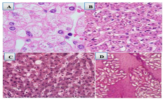
This study aimed to investigate gross and histopathological liver lesions caused by Platyhelminthes in camels slaughtered at a meat processing facility in the Dynile District, Somalia. The investigations were conducted between April and October 2024. A total of 340 slaughtered camels were examined during the study. Among these, 200 camels (58.8%) were found to have liver lesions associated with Platyhelminthes based on both gross post-mortem examination and microscopic histopathological analysis. Gross pathological changes included hepatic swelling (34.5%), yellowish discoloration (27.5%), hemorrhagic lesions (11.5%), bile duct dilation (9.5%), necrosis (7.0%), cirrhosis (5.5%), and exudative lesions (4.5%). Histopathological analysis revealed frequent features such as tissue infiltration (25.5%), lymphocytic infiltration (18.1%), granuloma formation (13.5%), eosinophilic infiltrates (13.0%), necrosis (11.2%), abscess formation (8.3%), inflammation (7.1%), fibrosis (4.2%), and hepatocellular necrosis (2.4%). These findings highlight the pathological burden of platyhelminth infections in camels and underscore the potential zoonotic risks associated with their transmission.

This work is licensed under a Creative Commons Attribution 4.0 International License.
Copyright (c) 2025 Abdirahman Barre, Faez F. J. Abdullah, Shafii A. Mohamed, Hamza A. Hashi, Krishnan N. Balakrishnan, Hodan I. Nageye (Author)